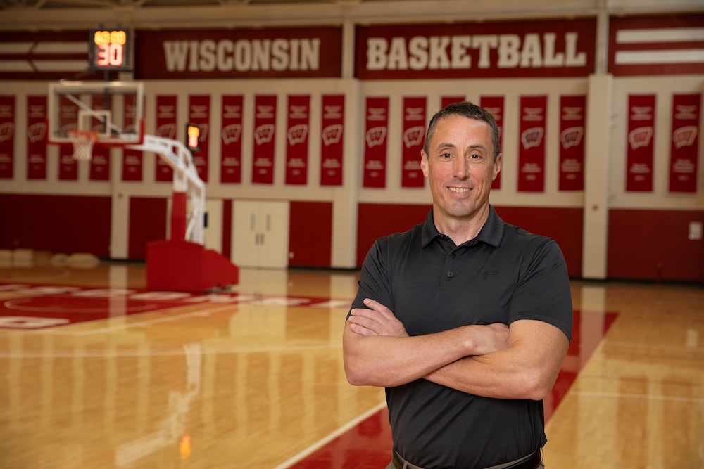
News & Events

See All News and Events

mRNA-activated blood clots could cushion the blow of osteoarthritis


Written by Tom Ziemer

University of Wisconsin-Madison researchers have developed a promising technique for treating osteoarthritis using therapeutic blood clots activated by messenger RNA.

Osteoarthritis is the most common form of arthritis, affecting roughly 33 million adults in the United States, according to the Centers for Disease Control and Prevention. It occurs when cartilage in key joints like the knees and hips deteriorates, causing pain and stiffness and impeding mobility.

Photo by Joel Hallberg

In a paper in the journal Bioactive Materials published online in December 2024, the UW-Madison research team led by William Murphy,PhD, a professor of biomedical engineering and orthopedics and rehabilitation, details its new approach. With further development, it could one day offer a more effective option than treatments such as steroid injections, hyaluronic acid injections or even joint replacement surgeries.

“The best-case scenario is that this could be an injectable or implantable treatment for patients who have advanced osteoarthritis,” says Murphy. “This would be an alternative to the existing methods for treatment, which generally don’t show a high level of long-term success.”

Following the lead of his lab’s previous work on mRNA-based vaccines, therapies for spinal cord injuries and more, the method relies upon mineral-coated microparticles to deliver mRNA that encodes for production of a protein that supports cartilage formation.

First, the team takes bone marrow aspirate (liquid bone marrow) and peripheral blood samples from a patient, mixes in the microparticles, and then forms the mixture into a blood clot. Then, the mRNA-activated clot gets delivered to the site of the damage.

“This all happens in the same surgery,” says Murphy, whose lab specializes in therapies that leverage biologically inspired materials. “This is all intra-operative, and it uses materials derived from the patient.”

Whereas existing treatments such as arthroscopic chondroplasties can lead to the formation of fresh fibrocartilage tissue, that material doesn’t boast the same mechanical properties of joint cartilage. It also degrades more quickly. Unlike traditional tissue engineering approaches, however, the new method doesn’t require the use of a synthetic scaffold material upon which to grow cells.

After seeing success in rabbit models, the group will test its treatment strategy in a larger animal model before proceeding toward human clinical trials.

Murphy says his group is also exploring the same approach to treat large skeletal muscle and bone defects.

William Murphy is the Harvey D. Spangler Professor of biomedical engineering and orthopedics and rehabilitation, as well as an H.I. Romnes Faculty Fellow and director of the Forward BIO Institute. Other UW-Madison authors on the paper include Brett Nemke, a research program manager in the School of Veterinary Medicine; Wan-Ju Li, an associate professor of biomedical engineering and orthopedics and rehabilitation; Mark Markel, a professor of medical sciences in the School of Veterinary Medicine; Yan Lu, a scientist in the School of Veterinary Medicine; Connie Chamberlain, a former scientist in the Murphy lab and current scientist in the Department of Surgery; Jae-Sung Lee, a former scientist in the Murphy lab and now an assistant professor at the University of Minnesota; Gianluca Fontana, a former scientist in the Murphy lab; MD-PhD student Joshua Choe; Hongli Jiao, an assistant scientist in Wan-Ju Li’s lab; and Michael Nelson and Margot Amitrano, graduate students in biomedical engineering.

Funding for the research came from a private gift from the Shannon family in support of the Musculoskeletal Regeneration Partnership, the National Institute on Aging (award number F30AG077748), and the UW-Madison Medical Scientist Training Program.


This story originally appeared on Engineering.wisc.edu

Special technique helps fix James’s clubfoot


With help from the UW Health Kids orthopedic surgery team, James, 5, is nearing completion of treatment for his condition. He’s also the kind of kid who doesn’t let something like clubfoot hold him back from playing sports and loving life.


With help from the UW Health Kids orthopedic surgery team, James, 5, is nearing completion of treatment for his condition. He’s also the kind of kid who doesn’t let something like clubfoot hold him back from playing sports and loving life.

“If you tell James he can’t do something, he will prove to you that he can,” says his mother, Heather Warpinski. “James taught himself how to swim when he was 3. He just didn’t like swimming lessons. He also played soccer last summer and is starting to get interested in football.”

The UW Health Kids orthopedic team treated James’ clubfoot with a widely accepted, essentially painless approach called the Ponseti Method, which involves five to eight weekly cast applications that gradually realign the tendons, ligaments, joint capsules, and bones in a child’s foot. Following the casting, the child wears a special set of boots and a brace until total correction is achieved.
.
.
Two UW Health physicians learned casting method from Dr. Ponseti

Few pediatric orthopedic teams throughout the nation can claim, as UW Health Kids can, that one or more of its doctors learned how to perform the Ponseti Method directly from its creator, the late Dr. Ignacio Ponseti.

Dr. Ken Noonan, a UW Health Kids orthopedic surgeon, trained with Dr. Ponseti in the early 1990s at the University of Iowa. Meanwhile, Dr. Blaise Nemeth, a UW Health Kids non-surgical orthopedic specialist, visited Dr. Ponseti on several instances to enhance his own mastery of the technique.

Before most orthopedic surgeons embraced the use of the Ponseti method, most children with clubfoot were treated with a more invasive surgery that essentially took the foot apart and put it back together – a procedure that frequently led to scarring and lifelong arthritis.

“Dr. Ponseti was essentially ignored by most orthopedic surgeons for decades,” Noonan says. “As word of the Ponseti Method began to spread online, families would come to Iowa City from all over the world to have their kids treated. Over the last 25 to 30 years, the Ponseti Method thankfully emerged as the standard form of treatment. Today, our entire team is trained [on] how to perform the Ponseti Method.”

About one in four children treated with the Ponseti Method require a “tune-up” surgery to help balance the muscles in the foot. James was one of these kids, and he has been doing well since Dr. Noonan performed the surgery in September 2024.

“We have been really happy with everything UW Health has done for James,” says Heather. “We got incredibly lucky that he found his way to us. He is an incredible kid and the light of our lives.”
.
..


This story originally appeared on uwhealth.org.

UW Researchers Forge New Path to Patellar Tendon Recovery


Patellar tendon injuries, common among athletes and active individuals, can derail lives with chronic pain and restricted mobility. Recognizing the significant negative impact these injuries have on the quality of life for those suffering from them, researchers at UW hope to revolutionize treatment and help affected patients reclaim their active lives. Led by Naoaki Ito, DPT, PhD, a postdoctoral researcher and Jack Martin, PhD, a research scientist, in the UW-Madison Department of Orthopedics and Rehabilitation and Badger Athletic Performance, and Scott Crawford, PhD, an assistant professor in the UW-Madison Departments of Kinesiology at the School of Education and Orthopedics and Rehabilitation, the team is taking an innovative approach to address the challenges of both treating and tracking recovery of the tendon.

Like a bridge, the patellar tendon connects the powerful quadriceps muscle to the tibia to enable smooth knee movement. When the tendon is injured, the standard treatment is exercise therapy. However, determining the optimal exercise load to promote healing is a major, ongoing challenge. Currently, clinical practices rely on external weight measurements to guide treatment, but these don’t tell the whole story, as patients’ bodies often compensate in subtle ways, masking the true load on the tendon.

Further, traditional imaging tools like ultrasound and MRI do not fully capture the tendon’s response to treatment, making it difficult to track recovery.

Game-Changers in Tendon Load Measurement and Recovery Tracking

Research studies led by Drs. Ito, Martin, and Crawford employ a novel approach to load measurement using a shear wave tensiometer – a wearable device co-developed by Dr. Martin here at UW that directly measures tendon load during exercise. Instead of relying on external weights, the team is developing a rehabilitation protocol guided by the actual load experienced by the tendon. This approach would give clinicians a clearer picture of what is happening inside the body, leading to more precise, individualized treatment and better healing outcomes.

Leveraging a long-standing research collaboration between the Department of Orthopedics and Rehabilitation and the Departments of Radiology and Medical Physics, Dr. Ito is working with Ivan Rosado-Mendez, PhD, Diego Hernando, PhD and Samuel Hurley, PhD to explore advanced imaging techniques to improve tendon health and recovery assessment. These techniques include diffusion tensor imaging (DTI), a form of MRI that measures the tendon’s microstructure, and quantitative ultrasound imaging techniques such as shear wave elastography and back-scatter anisotropy analysis, which can provide detailed insights into tendon properties – and offer a more accurate way to track tendon healing during rehabilitation.

Fueling Innovation Through Collaboration and Funding

In May 2024, Dr. Ito launched an ongoing study funded by the Department of Orthopedics and Rehabilitation Freedom of Movement Award to test the repeatability of DTI measurements in patients with patellar tendon injuries. Additionally, through matched funding from the UW-Madison Departments of Radiology and Medical Physics Radiology Research and Development Fund, Dr. Ito and his co-PIs are collecting quantitative ultrasound images to compare different imaging methods for assessing tendon health. And beginning in 2025, Drs. Ito, Martin, and Crawford will launch a new study funded by a UW Institute for Clinical & Translational Research (ICTR) pilot grant using shear wave tensiometry to measure tendon load during 25+ rehabilitation exercises. The team will also integrate quantitative ultrasound to explore the relationship between tendon load and tendon properties, leveraging additional funding from the Wisconsin Partnership Program (WPP) 20th Anniversary Postdoctoral Grant awarded to Dr. Ito in October 2024.

Transforming Patient lives

By integrating direct tendon load measurements using cutting-edge technology with advanced imaging techniques, the team aims to provide a more accurate and individualized approach to rehabilitation while offering valuable insights into patellar tendon healing and better tracking of a treatment’s effectiveness. “This novel line of research led by Dr. Ito has substantial promise in improving how we care for tendon injuries, leading to greater function and activity levels for our patients,” says Bryan Heiderscheit, PT, PhD, Frederick Gaenslen Professor and Vice Chair of Research for the Department of Orthopedics and Rehabilitation and Director of Badger Athletic Performance.

For countless patients, this research represents the potential for pain-free movement, restored mobility, and a renewed quality of life. With each discovery, Dr. Ito and his research partners are not just advancing science; they are offering hope to those sidelined by injury – and for anyone waiting to return to the activities they love, that hope is priceless.

David Goodspeed, MD, receives 2024 OTA Mentorship Award


Last month, the Orthopaedic Trauma Association (OTA) announced the 2024 OTA Award winners. Among the honorees was Dr. David Goodspeed, associate professor in the Department of Orthopedics and Rehabilitation at the University of Wisconsin School of Medicine and Public Health. Dr. Goodspeed earned the 2024 Mentorship Award for his contributions “to the clinical, academic, and professional growth of OTA member(s) via mentorship.”


Whether it’s teaching us how to reduce hips (on himself), taking the time to teach even when we’re (very) behind in clinic, or rounding with us at 9pm, Goody is always eager to contribute to our education. We are so grateful we get to learn from him everyday and we can’t think of anyone more deserving of this award!

– UW Orthopedic Surgery Residents

Congratulations, Dr. Goodspeed, on this well-deserved honor!

UW–Madison Researchers Receive $50,000 ICTR Pilot Award for Tendon Rehabilitation Study


Last month, the Institute for Clinical and Translational Research (ICTR) at UW–Madison announced the recipients of its 2024 Translational Basic and Clinical (TBC) pilot awards. Among the 11 projects funded is a study led by Jack Martin, PhD, a research scientist in the University of Wisconsin Department of Orthopedics and Rehabilitation, and Scott Crawford, PhD, an assistant professor in the University of Wisconsin Departments of Kinesiology at the School of Education and Orthopedics and Rehabilitation at the School of Medicine and Public Health. Their project, “Measuring Patellar Tendon Load during Exercise Using Wearable Sensors,” was awarded $50,000 to advance research on rehabilitation practices for tendon injuries.

Martin and Crawford’s study seeks to improve the accuracy of patellar tendon loading during rehabilitation after tendon injuries by:

  • Determining whether the external loading prescribed by physical therapists aligns with the actual load experienced by the patient’s patellar tendon;
  • Investigating the potential of electrical muscle stimulation to enhance tendon loading during rehabilitation exercises;
  • Exploring the use of quantitative ultrasound to monitor tendon health over time.

Co-researchers on the project include Naoaki Ito, PhD, a postdoctoral researcher in orthopedics and rehabilitation, and Ivan Rosado-Mendez, PhD, an assistant professor in medical physics and radiology. The proposal was accepted on September 27, 2024, with the awarded funds supporting this cutting-edge work to enhance patient recovery and outcomes in tendinopathy treatment.