News & Events

See All News and Events

UW-Madison Launches Study in Collaboration with NBA, NBPA, GE HealthCare, and Springbok Analytics to Advance Injury Reduction in Basketball


Researchers at the University of Wisconsin-Madison are leading a groundbreaking multi-institutional research study to better understand and reduce musculoskeletal injuries among high-level basketball players. The study, titled “Musculoskeletal Injury Profiling of Elite Basketball Players: A Framework for Injury Mitigation Strategies Through Integration of Biomechanics, Imaging and Data Analytics,” began in August 2025 and will run through July 2026.

This research is made possible through UW-Madison’s collaboration with the NBA, National Basketball Players Association (NBPA), GE HealthCare, and Springbok Analytics, which are collectively providing expertise and technology capabilities to support advanced athlete monitoring, leading-edge imaging analysis, funding, and additional resources.

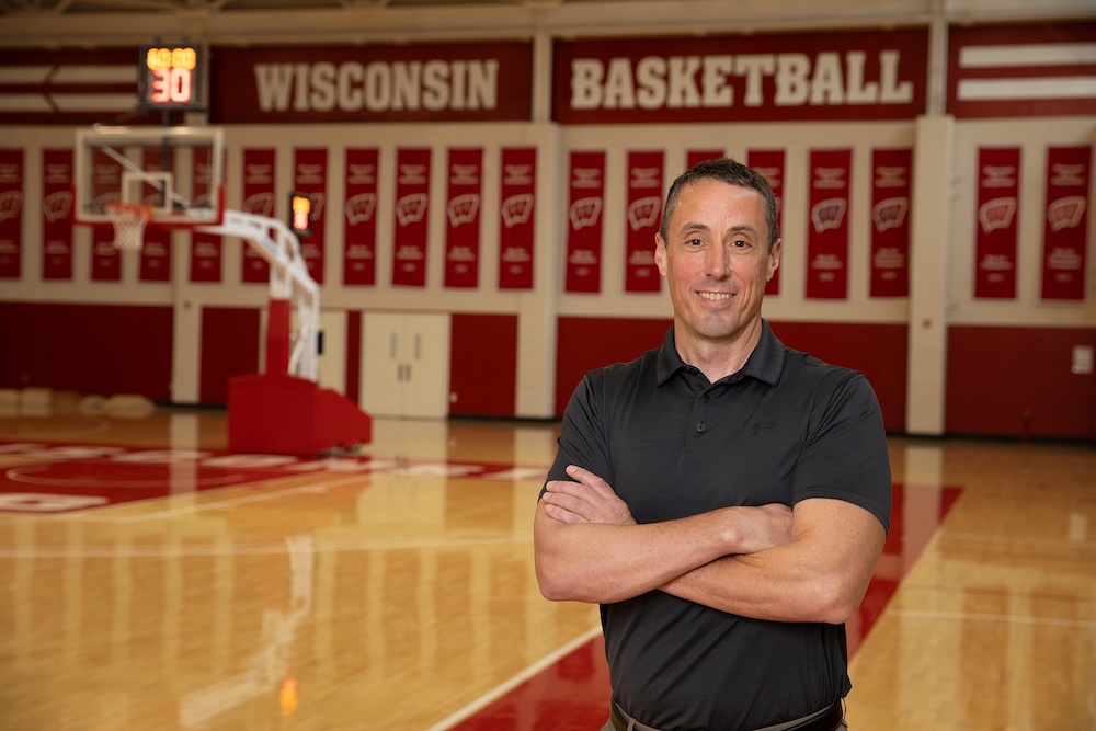
The project’s Principal Investigator is Dr. Bryan Heiderscheit, Frederick Gaenslen Professor in Orthopedics and Vice Chair for Research in the UW Department of Orthopedics and Rehabilitation, and Director of UW Badger Athletic Performance. The UW research team includes Drs. Matthew Blomquist, Naoaki Ito, Mikel Joachim, Kenneth Lee, and Jack Martin, and research coordinators, Alex Gruber and Claire Tanaka. Also critical to the project’s success are the teams’ athletic training and strength and conditioning staff, and the personnel at the additional research sites: Brigham Young University (BYU) and the Technical University of Munich, Germany.

The current study builds on the collaboration’s 2023-24 NBA G League pilot. The UW research team led the analysis of that pilot study, and the insights gained informed and expanded the scope of the present work. The current season-long study prospectively monitors potential risk factors for injury among collegiate and adolescent basketball players by leveraging movement biomechanics, magnetic resonance (MR) and quantitative ultrasound imaging techniques, strength assessments, and player training load. The study also utilizes Springbok’s 3D muscle volume quantification analysis of MR images to establish a detailed anatomical reference, enabling biomechanics, strength, and workload data to be mapped and analyzed over time in relation to injury and recovery. Before the competitive season, athletes participating in the study underwent a detailed data collection session to measure tendon and muscle characteristics and strength. Then, at key points during and at the end of the season, these assessments are repeated – with additional testing when injuries occur to track severity and recovery.

Ultimately, through these regular, detailed assessments, including the use of a novel ultrasound technique developed at UW to measure ankle ligament stability, Dr. Heiderscheit and his team aim to better understand factors contributing to common lower-body injuries and how athletes recover to full performance.

Freedom of Movement Awardee Andrew Watson, MD, aims to Improve Injury Prevention in Young Athletes


Andrew Watson, MD, associate professor in the UW Department of Orthopedics and Rehabilitation and director of the UW Human Performance Lab, has received a 2026 Freedom of Movement (FOM) Award in support of his research project, “The Interaction of Surface Type and Heat Stress to Influence Musculoskeletal Injury and Concussion Risk in Youth Athletes.” The award provides internal funding for a large-scale, data-driven study aimed at reducing injury risk in youth sports through evidence-based match scheduling.

Dr. Watson’s project addresses a critical gap in youth athletics. Although the risks associated with heat, playing surfaces, and competition demands are increasingly recognized, there is currently no evidence-based framework to guide match scheduling in ways that protect young athletes. This study will be among the first to prospectively combine real-world injury surveillance with detailed weather and playing surface data from elite youth soccer events across the United States, examining how heat stress, surface type, age, gender, and time of day interact to influence musculoskeletal injury and concussion risk.

A key strength of the project is its scale and real-world setting, made possible through a collaboration with the Elite Clubs National League (ECNL), the nation’s premier youth soccer development and competition platform, and the ECNL’s Center for Athlete Health and Performance. Dr. Watson’s lab has partnered with the ECNL for several years on initiatives related to athlete well-being, including mental health and injury prevention.

“Our collaboration with the ECNL,” said Dr. Watson, “has resulted in a fantastic opportunity to generate specific evidence that can translate into real-world interventions to benefit young athletes. This project builds on that relationship by advancing our understanding of how environmental factors influence injury risk and helping to develop a truly innovative approach to match scheduling that optimizes player health and safety.”

For this study, the ECNL is providing access to national injury surveillance data and match scheduling information. Certified athletic trainers at more than 40 ECNL national events will collect detailed injury information, allowing the research team to calculate precise injury rates across different age groups, genders, and playing surfaces. Data will be collected from events totaling an estimated 400,000 to 500,000 player-hours, providing the statistical power needed to identify meaningful injury patterns and interactions among multiple risk factors.

Injury surveillance data will be paired with historical local weather data to calculate wet bulb globe temperature, a standard measure of heat stress. This integrated approach will enable a more nuanced understanding of how environmental conditions influence injury risk in youth athletes.

The potential impact of this work is substantial. By examining how environmental and structural factors intersect, the research aims to identify previously unmeasured injury risk patterns and translate findings into practical, scalable match scheduling strategies that better protect young athletes while preserving competitive integrity. These findings could ultimately serve as a model for injury prevention, not only in youth soccer, but across a wide range of youth sports nationwide.

Collaborators on this project include Jennifer Sanfilippo, PhD, assistant director of sports medicine within UW Athletics and coordinator of Badger Athletic Performance, Christian Lavers, MBA, JD, president and CEO of the ECNL, and Kristin Haraldsdottir, PhD, a research scientist within the UW Human Performance Lab. Together, the team brings expertise in athletic performance, sports administration, injury epidemiology, and applied research, ensuring the project’s findings will be both rigorous and impactful.

“We are truly grateful to the UW Department of Orthopedics and Rehabilitation for the support of this ongoing work,” said Dr. Watson.

Eric Cotter, MD, receives Freedom of Movement Award for Research to Improve Outcomes after Reverse Shoulder Replacement


The UW Department of Orthopedics and Rehabilitation named orthopedic surgeon and assistant professor Eric Cotter, MD, a recipient of the 2026 Freedom of Movement (FOM) Award. Dr. Cotter’s FOM grant will support an innovative research project focused on improving outcomes after reverse total shoulder arthroplasty. The project, titled “Soft Tissue Tensioning in the Setting of Inlay, Lateralized Designed Reverse Total Shoulder Arthroplasty: A Cadaveric Study,” seeks to better understand how soft-tissue tension affects shoulder function following this commonly performed procedure.

Reverse total shoulder arthroplasty is now the most frequently performed type of shoulder replacement worldwide. Unlike a native shoulder joint, a reverse implant alters the normal ball-and-socket joint orientation, leading to changes in muscle mechanics and soft-tissue tension. While this approach has expanded surgical options for patients with complex shoulder conditions, debate continues surrounding how best to position implants and balance soft tissues to achieve optimal function.

Dr. Cotter’s study aims to define the ideal tension to apply during reverse shoulder replacement and how that tension affects surrounding structures. A novel aspect of the study is the use of a handheld tensiometer – a tool developed by co-investigator Josh Roth, PhD – to directly measure soft tissue forces within the shoulder during implantation. The project also incorporates a muscle–sparing surgical technique, visual markers to assess strain on the acromion as a surrogate for deltoid tension, and dynamic ultrasound imaging to evaluate shoulder joint stability.

The research team will conduct a cadaveric study of 20 shoulders, first simulating a standard clinical shoulder examination using ultrasound to establish baseline, native joint laxity. Reverse shoulder replacements will then be performed while systematically varying the thickness of the polyethylene component to incrementally increase soft-tissue tension. The team will collect measurements to assess how these changes influence acromial strain and tension within the conjoint tendon.

“This work,” says Dr. Cotter, “is one small step toward better understanding how tight we should be putting in these implants and if we can use a tensiometer for real-time feedback of soft tissue tension. This improved soft tissue balancing should lead to better long-term functional outcomes.”

Support from the Freedom of Movement Award enables this early phase of what Dr. Cotter hopes will be a larger research effort – one that has the potential to refine surgical decision-making in reverse shoulder arthroplasty in a way that optimizes postoperative range of motion for these surgical patients.

In addition to Drs. Cotter and Roth, Kyle Wagner, MD, Lauren Summers, MD, Andrew Sheean, MD, and Herman Feller, PhD, are co-researchers on the project. Pilot work is already underway, and the team anticipates completing the study by December 2026.

With clubfeet now straight, James is doing great


James Van Hierden now shows few signs of being born with clubfoot, a condition where one or both feet are twisted severely inward, after undergoing treatment by UW Health Kids experts.
.
.
James Van Hierden is among the one in 1,000 children born with clubfoot, a condition where babies enter the world with one or both feet twisted severely inward.


Babies who are born with clubfoot have a shorter Achilles tendon. This abnormality pulls the foot out of position.

James’s parents, Michael and Michaela Van Hierden, learned about their baby’s diagnosis during Michaela’s 20-week ultrasound.

Like any couple, the Van Hierdens were not prepared for this kind of news, but once James was born and began treatment with the UW Health Kids orthopedic surgery team, their stress transformed into trust that their son was in the right place for care.

“We weren’t familiar with clubfoot, so it was pretty scary at first,” Michaela said. “The pediatric orthopedic doctors assured us James would ultimately be fine.

“Now, 4 years old, his treatment is over and he’s doing great.”
.
.
Experts in painless Ponseti method

The pediatric orthopedic team treated James with a widely accepted, essentially painless approach called the Ponseti method. The clubfoot treatment involves weekly casting applications that gradually realign the child’s tendons, ligaments, joint capsules and bones, resulting in normal foot positioning.

Children typically need five to seven weekly castings. James required seven because his left foot was turned extremely inward.

Following the casting, a second phase of treatment requires the child to wear a special boots-and-bar brace that keeps the child’s feet in the corrected position while preventing a relapse. At first, the child wears the brace 23 hours a day, eventually tapering down to only wearing the brace overnight until treatment is complete, typically by age 4 or 5.

Dr. Lindsey Boyke, a UW Health Kids orthopedic specialist who cared for James, says the beauty of the Ponseti method is that it causes minimal distress to the child and, through casting, corrects three of the four abnormalities that comprise clubfoot.

The fourth abnormality is typically corrected through a surgical procedure called a tenotomy, which involves cutting the Achilles tendon so it will grow on its own to normal length. This eventually lets the ankle move more freely and heal in proper position.

James required a tenotomy on both feet, which was performed when he was approximately 4 months old.


James Van Hierden and Dr. Lindsey Boyke


Before most orthopedic surgeons embraced the Ponseti method, most children with clubfoot were treated with a far more invasive procedure that frequently led to recurrence, chronic pain and lifelong arthritis.

Over the past few decades, the Ponseti method has emerged as the gold standard for treating clubfoot.

Dr. Kenneth Noonan, one of Dr. Boyke’s UW Health Kids orthopedic colleagues, trained with the procedure’s namesake, Dr. Ignacio Ponseti, in the early 1990s at the University of Iowa. Another UW Health Kids orthopedic specialist, Dr. Blaise Nemeth, had the opportunity to meet and learn from Dr. Ponseti.

Today, the entire UW Health Kids orthopedic team is trained in the Ponseti method. James received care from all members of the team.

“We’re very lucky that our team has had so much first-hand exposure to Dr. Ponseti,” Dr. Boyke said. “Many of our newer faculty, including myself, learned the Ponseti method from Dr. Noonan and Dr. Nemeth.”
.
.
James thrives physically after treatment

Today, James is thriving with no restrictions on his activities.

“He’s always been surprisingly strong, even as a baby,” Michael said. “He hit milestones early, walking and running, and just kept going from there.

“Honestly, that’s meant a lot to us because we know what he might have faced without treatment. Seeing him thrive physically has been incredible.”

Both of James’ parents are health care professionals. Their experience has provided them a unique appreciation for the care their son received from UW Health Kids.

“Health care is navigating some significant challenges right now, and as clinicians and now parents, we’ve come to deeply value programs like this,” Michaela said.

“We hope to offer encouragement to other families, many of whom travel long distances for this kind of care. We always felt so well cared for and the team always made sure we had everything we needed to care for James.”
.
..


This story originally appeared on uwhealth.org.

Naoaki Ito, DPT, PhD, awarded Five-Year NIH Research Grant


Last month, Naoaki Ito, DPT, PhD, a postdoctoral researcher in the UW Department of Orthopedics and Rehabilitation and Badger Athletic Performance, was awarded a five-year, $945,000 K99/R00 Pathway to Independence Award from the National Institutes of Health (NIH) to fund his research on improving outcomes following patellar tendon injuries. This highly competitive grant supports early-career scientists in transitioning from mentored postdoctoral research to independent academic positions. Dr. Ito’s project begins with a two-year mentored phase, followed by three years of independent research as a tenure-track investigator.

With support from his K99/R00 award, Dr. Ito and his team are developing a new approach to optimize treatment for two common knee injuries: patellar tendinopathy (often known as jumper’s knee) and secondary injuries from ACL surgery that uses the patellar tendon as a graft, which can significantly impair mobility and quality of life. The team’s work focuses on better understanding how the tendon responds to exercise-based rehab – the gold standard treatment – and creating more precise and personalized treatment strategies.


This prestigious recognition highlights Dr. Ito’s exemplary line of research as an early career clinician-scientist focused on advancing strategies to restore and maintain tendon health.

Bryan Heiderscheit, PT, PhD
Frederick Gaesnlen Professor in Orthopedics
Vice Chair of Research
Department of Orthopedics and Rehabilitation
University of Wisconsin School of Medicine and Public Health


Dr. Ito’s primary mentors during the two-year mentored phase include:

  • Bryan Heiderscheit, PT, PhD, FAPTA, Frederick Gaenslen Professor and Vice Chair of Research in the Department of Orthopedics and Rehabilitation who will leverage his extensive experience in managing large-scale clinical research studies to provide Dr. Ito guidance on managing his first clinical trial; and
  • Darryl Thelen, PhD, the John Bollinger Chair of Mechanical Engineering and Bernard A and Frances M Weidman Professor in the UW College of Engineering and the inventor of shear wave tensiometry who will contribute his expertise for implementation of the technology in a clinical setting; and
  • Diego Hernando, PhD, Associate Professor of Radiology and Medical Physics at UW and a leader in the field of diffusion-weighted imaging will mentor Dr. Ito on the novel implementation of diffusion tensor imaging in tendons.

Dr. Ito will also be supported by co-mentors:

  • Stephanie Kliethermes, PhD, Associate Professor in the UW Department of Orthopedics and Rehabilitation and Research Director of the American Medical Society for Sports Medicine, who will bring her expertise in biostatistics and clinical trial design; and
  • Karin Silbernagel, PT, PhD, FAPTA, Professor and Associate Chair of the Department of Physical Therapy at the University of Delaware who is a world-renowned leader in tendon rehabilitation.

In addition, collaborators Samuel Hurley, PhD, assistant professor in the UW Department of Radiology, and Jack Martin, PhD, a research scientist in the UW Department of Orthopedics and Rehabilitation, will provide further support in the application of imaging and tensiometry techniques.

Dr. Ito’s research incorporates two emerging technologies to better understand how tendons respond to rehabilitation. One is shear wave tensiometry, using a wearable device capable of accurately measuring tendon force during movement and exercise – something that has been difficult to quantify in the past. The other is diffusion tensor imaging, an advanced MRI technique that provides detailed insight into the internal structure of tendons, allowing researchers to track tissue changes and recovery over time.

In addition to these tools, Dr. Ito’s team is evaluating whether neuromuscular electrical stimulation (NMES) – which uses electrical pulses to activate muscles – can enhance tendon loading during rehab and improve healing outcomes. By combining these innovative technologies with rigorous clinical research, the project aims to create more effective rehabilitation protocols tailored to individual patient needs.

Ultimately, Dr. Ito hopes this work will not only improve treatment for knee injuries but also pave the way for better approaches to tendon rehabilitation across the body – and smarter, science-driven strategies that help patients recover more fully and return to their daily activities with confidence.